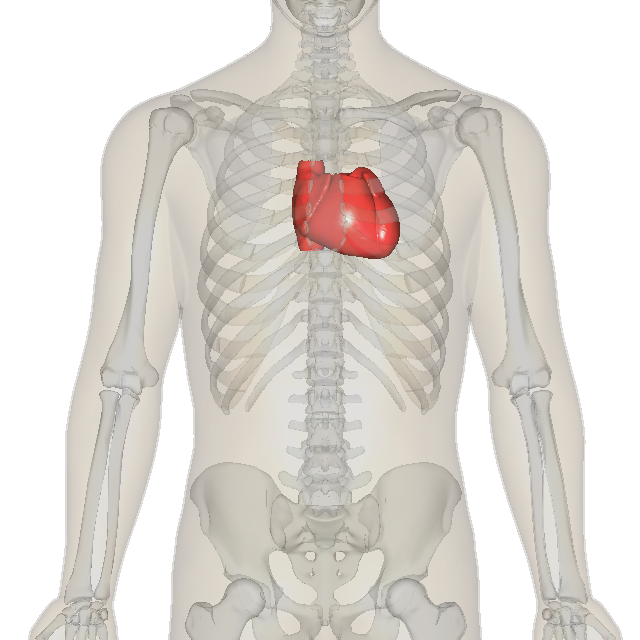
The human heart is a muscular pump, roughly the size of a clenched fist, that drives blood through the body every moment of life. Beating around 100,000 times a day and some 2.5 billion times over an average lifetime, it is one of the hardest-working organs in the body, and it never rests.
The heart is divided into four chambers: two upper chambers called atria and two lower chambers called ventricles. The right side receives oxygen-poor blood returning from the body and pumps it to the lungs; the left side receives oxygen-rich blood from the lungs and pumps it out to the rest of the body. A set of one-way valves between the chambers keeps blood flowing in the correct direction, and the familiar "lub-dub" of a heartbeat is the sound of these valves snapping shut.
The heart is one of the first organs to form and function in a developing embryo, beginning to beat just weeks after conception, long before most other organs have taken shape. From that moment it does not stop, beating ceaselessly for the whole of a person's life.
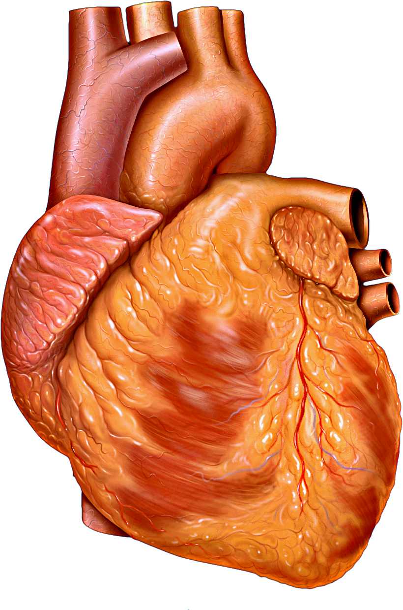
The heart is made largely of a special kind of muscle, the myocardium, found nowhere else in the body. Unlike the muscles we move at will, it contracts tirelessly and automatically, and it is richly supplied with its own blood vessels, the coronary arteries, to feed its relentless work.
The heart powers two linked circuits. In the first, it sends blood the short distance to the lungs to pick up oxygen and release carbon dioxide. In the second, it pumps that oxygen-rich blood out through the arteries to every tissue in the body, from which it returns, depleted, through the veins. This double circulation ensures that the body is supplied with the oxygen and nutrients it needs while waste is carried away.
Remarkably, the heart sets its own rhythm. A small cluster of specialised cells, the sinoatrial node, acts as a natural pacemaker, sending out electrical impulses that spread through the heart muscle and trigger each coordinated contraction. This is why a heart can keep beating on its own, and it is the activity that an electrocardiogram, or ECG, records.
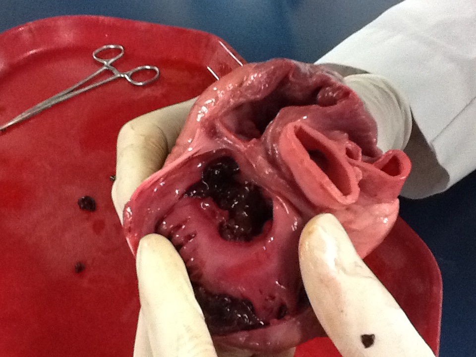
The two sides of the heart are not built the same. The left ventricle, which must push blood all the way around the body, has a far thicker, more powerful muscular wall than the right, which only sends blood the short way to the lungs. This difference is clearly visible when the heart is cut open.
Heart disease is among the leading causes of death worldwide, often the result of arteries narrowed by fatty deposits that can starve the heart muscle and cause a heart attack. Much of this risk is influenced by how we live: diet, exercise, blood pressure, and smoking all play a part. The heart's tireless, lifelong work makes its care one of the most important factors in human health.